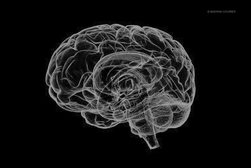
The human brain is essentially a ball of meat that’s charged with electricity. There are neurons, fibres, and synapses that keep it going. There are some popular parts, like the pre-frontal cortex, and some not-so-popular parts, like the Wernicke area. When the brain starts to malfunction, the entire body suffers. Brain mapping, a scientific way of scanning the brain, can prevent that.
Brain mapping, essentially, is creating a map of the activities occurring in the brain by scanning and analysing the electrical activity in the brain. It can be used to treat addictions and diseases like brain strokes, seizures and brain tumours. Researchers use laser stimulation techniques and magnetic resonance imaging (MRI) to create an image of the brain.
After imaging the brain by the laser technique, the brain is mapped using a connectogram or a graphical representation. This representation depends on the graph theory. Graph theory, essentially, is the combination of points and lines that connect these points. They are different from charts like bar graphs and pie charts. The focus is merely on lines and points.
Brain mapping began in the late 1970s when MRI and CT scans were invented. These two ways of brain mapping were much less harmful than the others that were available at the time. Today, the MRI scan is the most popular way of scanning the brain.
MRI scanning uses a magnetic field and computer-generated radio waves to take images of the body. It is more accurate, more clear than X-rays. When a patient goes inside the MRI scanner, the magnetic fields rearrange the water molecules in the body, and the radio waves come in contact with these molecules, causing them to produce signals which are used to create the image in the scanner.
It is important not to have any metal on the body while doing the MRI scan because that could lead to dire consequences.
Another way to map the brain is to get an electroencephalogram (EEG) scan. An EEG is like a screenshot of the brain. There are small metal discs that are attached to the scalp. While the metal discs are attached, the brain’s activity is recorded as wavy lines. It can also be used to see if someone is in a coma or is dead. However, one has to be very careful about this since sometimes EEG causes seizures if a patient has a seizure problem.
Apart from these two, there are other ways of scanning the brain, like positron emission tomography (PET), electrocorticography (ECoG), magnetoencephalography (MEG), and optical imaging with near-infrared spectroscopy (NIRS).
Although the techniques used are relatively new, the ideas behind them are old. For example, the idea that brain activity changes through blood flow, which can be observed, came about in the nineteenth century. An Italian physiologist, Angelo Mosso, observed the brain pulsations while the patients did tasks like mathematics. The blood flow increased When they did tasks requiring the brain to work harder. Later, two gentlemen, Charles Roy and Charles Sherrington, established a connection between brain activity and blood flow.
Later, many scientists came in and proved that blood circulation in the brain is related to activities that occur in the brain, thus effectively proving that mapping the brain’s activity can tell us the causes and treatments of the problems inside the brain. Yet up until 1986, it was believed that the reason for the increase in blood flow was the need for more oxygen, which points out to biological stimulation instead of external.
But slowly, with the emergence of CT and MRI, it was found that the connection between blood flow and activity in the brain depends on an external phenomenon like doing maths or drinking alcohol than biological. That gave impetus to the practice of brain mapping.
Another reason why brain mapping is used is so that neurosurgeons can be cautious in their practice. They must ensure they are not touching a sensitive area when performing surgery. Looking at the brain’s activity beforehand can determine the problem, what has to be done, and how it should be done.
Brain mapping is essential to those with a brain tumour, especially when the doctor has to ensure that other parts of the brain are not affected while removing the tumour. These other parts could include motor pathways and the brain area for languages.
A neurosurgeon has to be very careful in treating the tumour. Brian mapping ensures that the neurosurgeon knows exactly where the tumour is and how to treat it.
To sum it up, brain mapping is vital to know what caused the damage in the brain and how it can be treated or prevented. No matter what method is used for the procedure, one must always know what one can do to be fully prepared. If one is not able to know, at least their near and dear ones should be aware.
-30-
Copyright©Madras Courier, All Rights Reserved. You may share using our article tools. Please don't cut articles from madrascourier.com and redistribute by email, post to the web, mobile phone or social media.Please send in your feed back and comments to [email protected]